




BLOG > mRNA & LNP > LNP-mediated mRNA delivery in human PBMC: T cell-targeted transfection
T Cell Transfection - Application Note
In collaboration with CARMIL Therapeutics & JC Discovery, we evaluated an LNP formulation dedicated to T cells using flow cytometry analysis.
Background & Objective
Lipid Nanoparticles (LNPs) represent a highly efficient non-viral delivery system for messenger RNA (mRNA), enabling transient protein expression in a broad range of cell types. OZ Biosciences has formulated a specific LNP formulation based on new proprietary ionizable lipid encapsulating an eGFP-encoding mRNA as a fluorescent reporter model.
The objective of this first-in-series study, conducted in partnership with CARMIL Therapeutics & JC Discovery, was to evaluate the transfection efficiency, cell-type tropism and cytotoxicity of this LNP formulation in human peripheral blood mononuclear cells (PBMCs), aiming more specifically at transfecting T cell subset of this therapeutically relevant primary cell model.
Experimental Design
Human PBMC were sourced from frozen donor samples and pre-activated for 48 hours with anti-CD3/CD28 antibodies prior to LNP treatment. GFP mRNA (5’-Cap1, N1-methyl-pseudouridine, Poly(A)-3’) was encapsulated in novel ionizable lipid LNP formulation.
Key Parameters:
Doses: 0.5 & 1.5 µg | Cell density: 2.5 x 10⁵ cells/conditions | Incubation: 20 hours.
Cell Viability
Cell viability was assessed by flow cytometry for both T cells and total leukocytes across all doses. As shown in Figure 1, LNP treatment did not induce significant cytotoxicity even at the higher dose. Viability remained above 85% in all conditions in both T cells and leukocytes, comparable to untreated control (No LNP), demonstrating excellent biocompatibility of the LNP formulation.
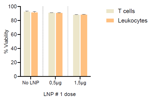
Figure 1: Cell viability (%) of T cells and total leukocytes following LNP treatment at 0.5 and 1.5 µg. Data shown as mean ± SEM (duplicates). No LNP: untreated control.
Transfection Efficiency
Expression gating
Flow cytometry dot plots show the proportion of GFP-positive cells detected within each immune cell subpopulation (T cells, CD4+ T cells, CD8+ T cells, B cells, NK cells and NKT cells), under three conditions: No LNP, 0.5 µg and 1.5 µg of LNP formulation.
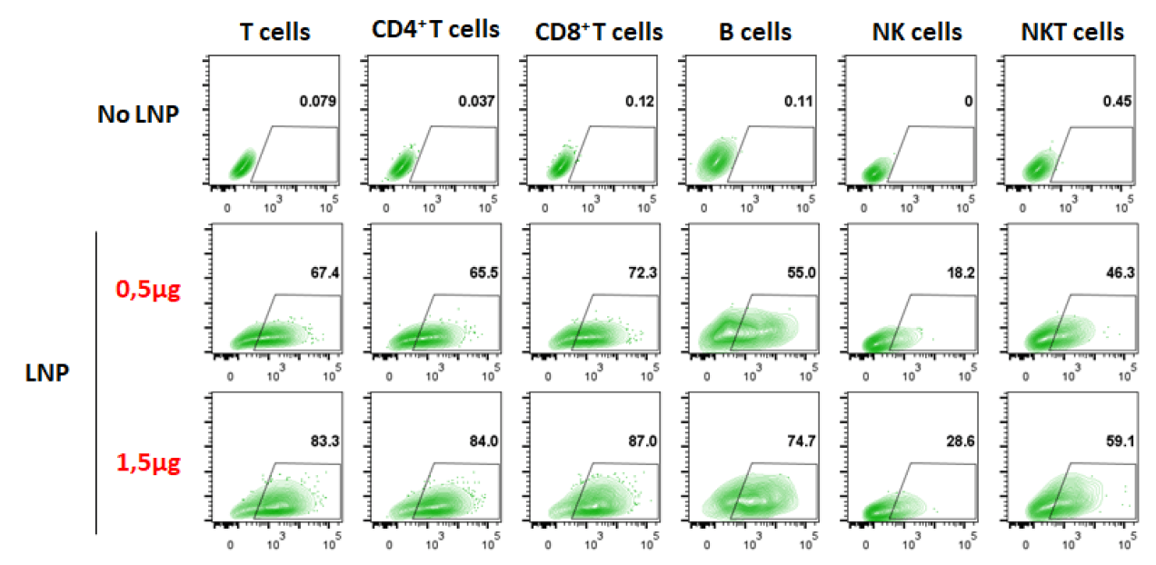
Figure 2: Representative flow cytometry dot plot of GFP expression across immune cell subpopulations. Values indicate % GFP+ cells within gated population.
GFP+ cells as a percentage of total leukocytes
When expressed as a proportion of total leukocytes, T cells emerged as the predominant transfected population, reaching approximately 57% at 0.5 µg and 69% at 1.5 µg LNP. Within T cell compartment, both CD4+ and CD8+ subsets contributed equally at around 25-31%. B cells, NK cells, and NKT cells showed markedly lower transfection from total leukocytes, reflecting the T cell propensity of this LNP formulation over other cell types under activated PBMC conditions.
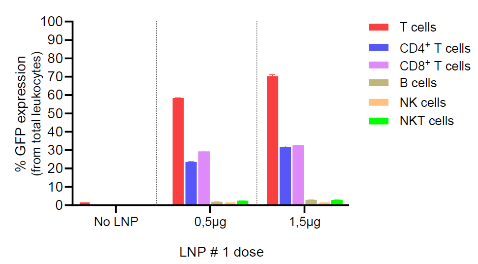
Figure 3: Percentage of GFP+ cells from total leukocytes following LNP treatment. Color-coded by cell subpopulation.
GFP+ cells within each cell subpopulation (intrinsic transfection rate) Normalizing GFP expression within each cell subpopulation independently reveals a strikingly high intrinsic transfection rate across all immune cell types. At 1.5 µg LNP, T cells (83.3%), CD4+ T cells (84.0%), CD8+ T cells (87%) showed near-complete transfection. B cells also achieved high expression (74.7%) while NKT cells and NK cells reached 59.1% and 28.6% respectively. These results confirm that LNP formulation efficiently transfect the vast majority of pre-activated T and B lymphocytes and a substantial fraction of innate immune cells.
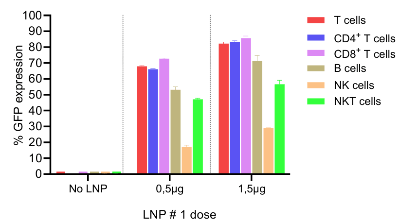
Figure 4: Percentage of GFP+ cells within each individual cell subpopulation following LNP formulation treatment at 0.5 and 1.5 µg.
Summary Table: GFP mRNA Delivery at 1.5 µg with LNP formulation
Table 1 below compares the two LNP formulations head-to-head at the highest tested dose (1.5 µg), showing % of GFP+ cells within each cell subpopulation. Background fluorescence (No LNP condition) ranged from 0.08% to 0.45% across all populations.
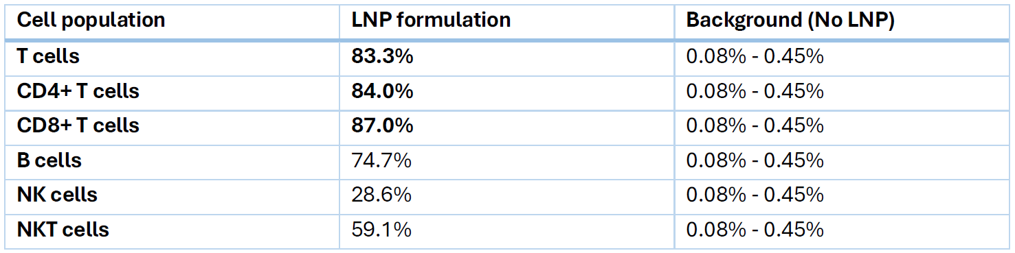
Table 1: Summary of GFP+ cell percentage within each immune cell subpopulation for LNP formulation. Data collected by CARMIL Therapeutics and JC Discovery.
Conclusion
This first evaluation of our LNP formulation in human primary PBMCs demonstrates a strong proof of concept for LNP-mediated delivery in therapeutically relevant immune cell populations. Our new ionizable lipid LNP formulation achieves exceptionally high transfection rate in pre-activated T cells and B cells with no detectable cytotoxicity, making it a highly promising candidate for further development.
We want to warmly thank CARMIL Therapeutics for their collaboration in this study.
This formulation is already available upon request and will be added to our catalog soon, stay tuned for more details.
Related publications
Stem cell mRNA transfection: LNP delivery Application Note
White paper: Ionizable lipids for mRNA-LNP and siRNA-LNP delivery