



BLOG > Publications & Citations > Development of a gene-activated matrix for enhanced AAV gene delivery in vitro
From Scaffold to gene delivery : How a gene-activated matrix combined with AAVBlast enables efficient local AAV-mediated bone regeneration in vitro
Gene therapy for bone regeneration holds great promise, but delivering therapeutic genes precisely where they are needed without systemic exposure remains a key challenge. When critical-size bone defect fails to heal, current gold-standard treatments carry significant risks and limitations. Musoki et al. tackled this challenge by developing a gene-activated matrix (GAM) combining chitosan/β-tricalcium phosphate scaffolds with AAV2 vectors, and by identifying AAVBlast as a novel poloxamer-based transduction enhancer, ultimately achieving localized delivery of osteogenic and angiogenic growth factor in vitro.
Key results
The study delivered several interconnected findings that together establish a robust GAM platform:
Among AAV2, AAV6 and AAV8, AAV2 was identified as the superior serotype across all tested cell types, with fluorescent levels almost 8-fold higher than AAV6 in Hela cells and 5-fold higher in primary ovine mesenchymal stromal cells (oMSC). All subsequent experiments were conducted based on this observation.
Incorporation of β-tricalcium phosphate (β-TCP) into chitosan scaffolds significantly reduced AAV release from 15.7% (pure chitosan) to ~6.6% (TCP-containing scaffolds), indicating enhanced vector retention within the matrix. Scaffolds containing 10% β-TCP were selected as the optimal formulation, balancing retention, release, and cell attachment.
oMSC seeded onto AAV2-loaded GAMs produced BMP-2 and VEGF confirming efficient in situ transduction.
AAVBlast was identified as the most potent transduction enhancer delivering 6.4-fold increase in fluorescence at the lowest concentration tested in oMSC. AAVBlast stabilized AAV particles, elevated intracellular AAV DNA levels, transgene mRNA expression and protein production across HeLa, HEK293T, HUVEC and oMSC.
Modular application of AAVBlast onto the GAM increased detectable released vector genomes 4.2-fold at 6h, and significantly boosted BMP-2 production and VEGF secretion in oMSC.
Methodology: a modular, stepwise development strategy
The workflow was built sequentially, allowing each component to be independently validated before integration into the full GAM system:
- AAV serotype and transgene format selection was the first step. dsAAV2, dsAAV6 and dsAAV8 encoding eGFP were screened across HeLa, HEK293T, Saos-2; and primary oMSC. Single-stranded (ss) and double-stranded (ds) DNA formats were then compared for BMP-2 and VEGF expression in oMSC, identifying ssBMP-2 and dsVEGF as optimal configurations.
- Chitosan/β-TCP scaffold fabrication involved freeze-drying at -80°C and then -100°C under vacuum, yielding 90% porosity with pore size of 20-200 µm. Four TCP concentrations (0%, 10%, 20% & 30%) were evaluated for AAV release kinetics by qPCR and for in situ transduction efficiency by oMSC seeding.
- GAM generation and characterization involved direct loading of AAV2 onto scaffolds, followed by oMSC seeding and 3-week culture. BMP-2 and VEGF were quantified by ELISA and normalized to cell metabolic activity.
- AAVBlast screening was performed across multiple poloxamer formulations on primary oMSC using reporter gene-expressing AAV2. Mechanistic characterization included intracellular AAV DNA quantification, RT-qPCR, protein production assay and capsid ELISA for stability assessment.
- Osteogenic gene expression was analyzed at 4 weeks post-transduction by RT-qPCR in oMSC transduced with BMP-2 abd VEGF-expressing AAV2 vectors, with or without AAVBlast.
- Macrophage transduction was assessed by flow cytometry in primary monocyte-derived macrophages (MDMs) transduced with eGFP-expressing AAV2 in the presence or absence of AAVBlast.
AAVBlast in this study
AAVBlast was applied at 0.5-1 µL per well (96-well format, final concentration ~0.4%), prepared in serum-free medium on ice and combined with AAV vector prior to cell addition, or applied sequentially onto AAV-loaded scaffolds with a 10-minute gelation step at 37°C before oMSC seeding.
A screening of 6 poloxamer formulations on primary oMSC using reporter gene-expressing AAV2 identified AAVBlast as the clear winner, significantly outperforming all other candidates including F68 as well as the reference enhancer MG-132 (Figure 3A). The enhancement was not cell-type specific: similar gains were observed across HeLa, Saos-2 and HUVEC cell lines (Figure 3B), and across AAV serotypes 2, 6 and 8 (Figure 3C), confirming broad spectrum activity of AAVBlast.
Although the precise mechanisms of action of AAVBlast have not yet been fully elucidated, they likely involved the amphiphilic properties of its poloxamer-based components. The hydrophobic core of poloxamer unimers can insert into plasma membrane lipid bilayers, decreasing membrane viscosity and increasing lipid exchange, a membrane remodeling effect known to facilitate viral vector entry. In addition, the cationic nature of AAVBlast is expected to reduce electrostatic repulsion between negatively charged AAV capsids and the cell surface, thereby enhancing vector adsorption and uptake.
To assess its mechanism of action, AAV2 was incubated at 37°C in the presence or absence of AAVBlast. AAVBlast significantly increased detectable AAV2 capsid levels by 6.2-fold compared to untreated controls (Figure 3D), indicating enhanced particle stability and bioavailability. Consistent with these findings, AAVBlast treatment resulted in significantly elevated intracellular AAV genome copy numbers (Figure 3E), transgene mRNA expression (Figure 3F), and eGFP-mediated fluorescence (Figure 3G) across HEK293T, Saos-2, and primary oMSC; demonstrating that AAVBlast acts at multiple levels of the transduction process.
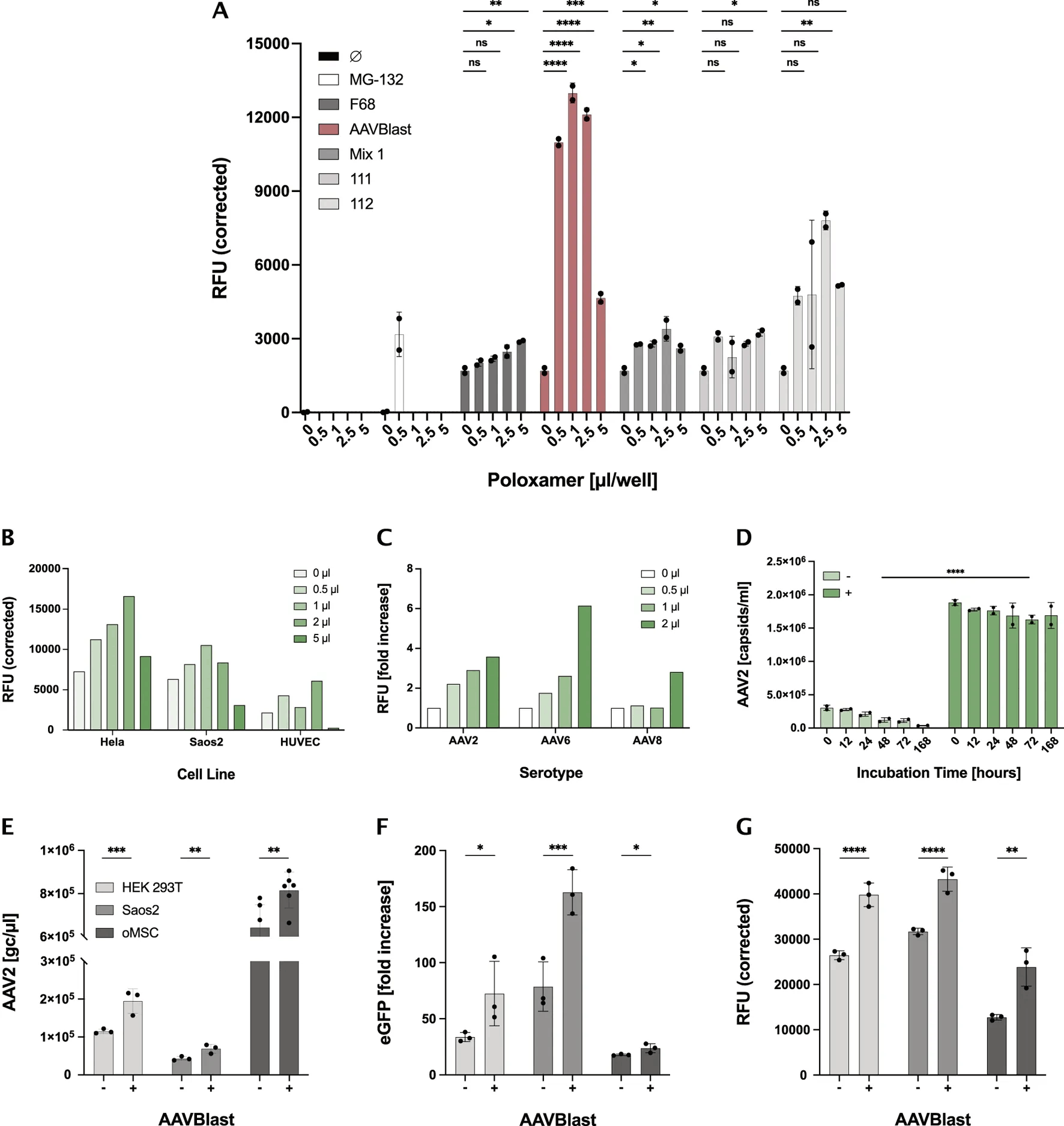
Figure 3: Poloxamer AAVBlast enhances AAV-mediated gene transfer. (A) AAVBlast was identified as the most potent among all poloxamers screened on primary oMSC, significantly outperforming MG-132 at optimal dose. (B) Enhancement confirmed across HeLa, Saos-2 and HUVEC. (C) Effect validated across AAV2, AAV6 and AAV8 serotypes. (D) AAVBlast increased detectable AAV2 capsids 6.2 fold after incubation at 37°C. (E-G) Significant increase in intracellular AAV2 DNA, transgene mRNA and eGFP fluorescence across multiple cell types. Adapted from Musoski et al., Front. Bioeng. Biotechnol. 2026, 14:1832901
Beyond transduction efficiency, AAVBlast also demonstrated a meaningful impact on osteogenic gene expression in primary oMSC transduced with BMP-2 and VEGF expressing AAV2 vectors, assessed by 4 weeks post-transduction. AAVBlast significantly enhanced BMP-2 mRNA expression in cells co-transduced with both vectors (Figure 4A), and increased VEGF mRNA levels ~2.5 fold in VEGF-transduced oMSC (Figure 4B). Interestingly, while early osteogenic markers Runx2 and Osterix were reduced by AAVBlast at this time point (Figures 4C, D), the late osteogenic markers Osteocalcin and Osteopontin were significantly upregulated (Figures 4E, F), suggesting that AAVBlast promotes more advanced stages of osteogenic differentiation driven by AAV-delivered growth factor signaling.
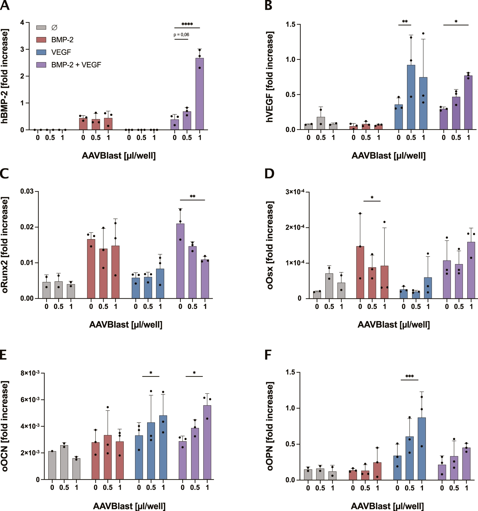
Figure 4: AAVBlast promotes AAV-mediated osteogenic gene expression. Primary oMSC were transduced with AAV2-BMP-2 and AAV-VEGF, separately or in combination, and gene expression was analyzed at 4-weeks post-transduction. AAVBlast significantly enhanced transgene expression of (A) BMP-2 and (B) VEGF. While early markers (C) Runx2 and (D) Osteorix were reduced at this late time point, the late osteogenic markers (E) Osteocalcin and (F) Osteopontin were significantly increased in the presence of AAVBlast. Adapted from Musoski et al., Front. Bioeng. Biotechnol. 2026, 14:1832901.
When applied onto the GAM, AAVBlast significantly increased detectable released AAV2 genome copies, with the most pronounced effect at 6h, rising 4.2 fold from 5.3 (±1.3)x105 to 2.2(±0.23)x106 genome copies (Figure 5A). This translated into significantly enhanced infectivity of released vectors, as confirmed by eGFP expression in HEK293T cells transduced with GAM-released AAV (Figure 5B). Importantly, in situ transduction by scaffold-retained AAV was comparable between AAVBlast-treated and untreated conditions (Figure 5C), suggesting that AAVBlast primarily stabilizes and protects released vectors rather than acting on scaffold binding.
Finally, application of 1µL AAVBlast onto growth factor-expressing GAMs significantly increased BMP-2 secretion 2.29-fold (Figure 5D), and VEGF secretion (Figure 5E) in oMSC seeded on the scaffold after 3 weeks.
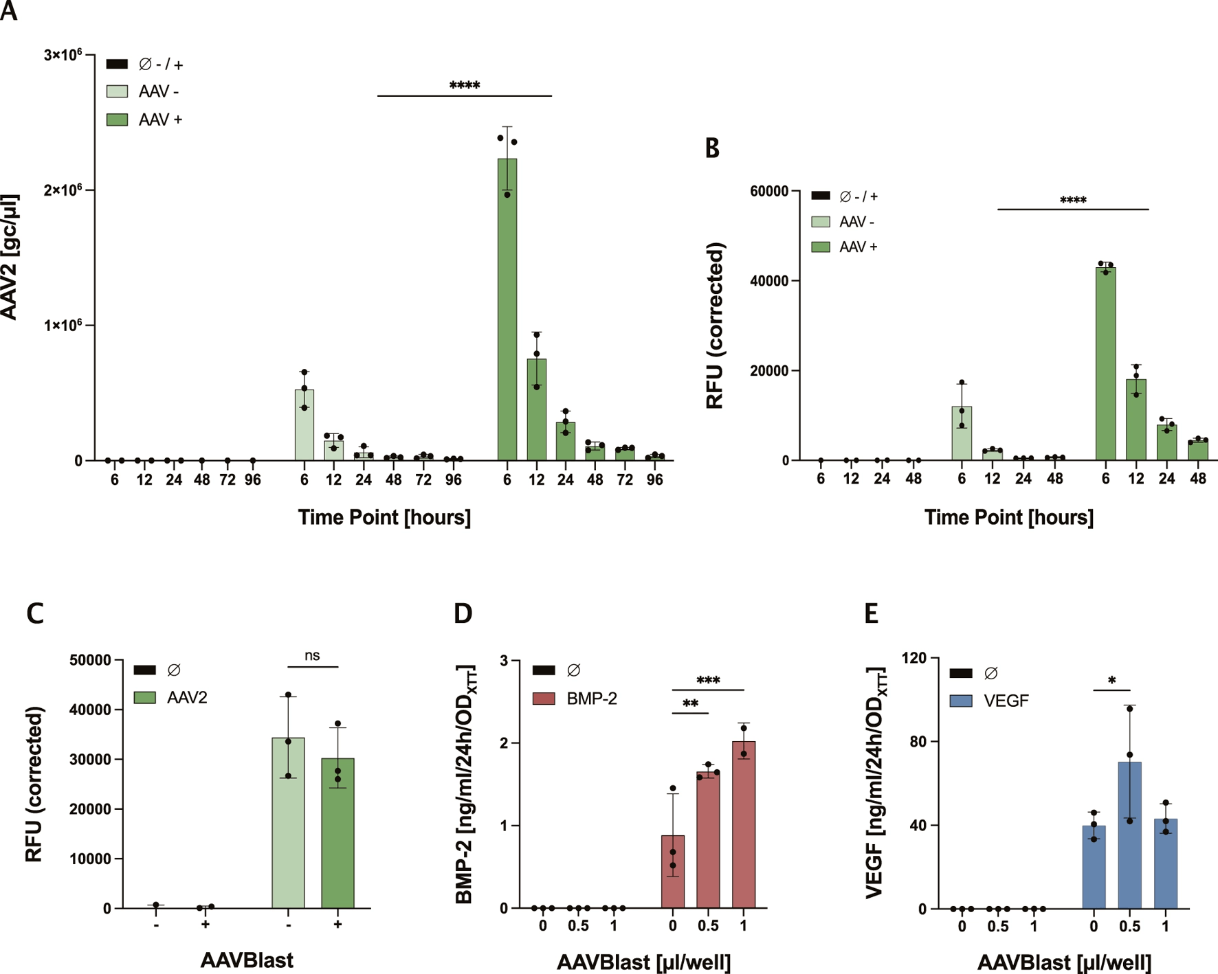
Figure 5: Poloxamer AAVBlast enhances transduction of Gene-activated Matrix-released AAV2. (A) AAVBlast significantly increased detectable released AAV2 genome copies. (B) Infectivity of released vectors significantly enhanced in HEK293T cells. (C) Scaffold-retained AAV transduction of oMSC was comparable with or without AAVBlast, indicating AAVBlast primarily acts on released particles. (D,E) BMP-2 and VEGF secretion by oMSC seeded on AAVBlast-enhanced GAMs were significantly increased after 3 weeks. Adapted from Musoski et al., Front. Bioeng. Biotechnol. 2026, 14:1832901
The authors also highlight that co-treatment with proteasome inhibitor MG-132 showed synergistic enhancement with AAVBlast, pointing to complementary mechanisms at distinct steps of the AAV transduction pathway. AAVBlast is non-toxic, requires no additional equipment or cell surface manipulation, and can be applied modularly onto existing AAV-based workflows, making it a broadly applicable tool for AAV gene delivery, both in standard transduction settings and in matrix-based delivery systems.
SBR Grant Project – Horizon 2020
This work was conducted within the framework of the SBR (Smart Bone Regeneration) project, funded by the European Union’s Horizon 2020 research and innovation program under grant agreement No. 874896.
Bringing together academic and industrial partners across Europe, SBR aims to develop next-generation strategies for the treatment of critical-sized bone defects. This publication represents a key proof-of concept milestone within the project, establishing a modular, transferable GAM workflow that combines biomaterial engineering, AAV gene delivery, and transduction enhancement into a single integrated platform.
Reference: Musoski A, Poulhès F, Sapet C, Iqbal N, Ganguly P, Kampick M, Hoechst B, Marotta M, Dumler K, Jha A, Jones EA, Giannoudis PV, Blanchy M, Knolle P, Zelphati O and Anton M (2026) Development of a gene-activated matrix for enhanced AAV gene delivery in vitro. Front. Bioeng. Biotechnol. 14:1832901. doi:10.3389/fbioe.2026.1832901
Related Product: AAVBlast Transduction Enhancer
Related publications
Enhancing transduction efficiency of lentiviral vectors in CAR-T cell therapy
Challenges in AAV-Based Retinal Gene Therapies