




BLOG > Technologies & Applications > White paper: LentiBlast Premium, cationic poloxamers for lentiviral transduction
Enhancing gene transfer into hematopoietic stem cells at lower viral dose, while preserving cell viability.
This white paper examines the development and the achievement of LentiBlast Premium, a new class of patented cationic poloxamers engineered at OZ Biosciences to enhance lentiviral transduction using an example, hematopoietic stem cells. We present data demonstrating how its cationic block-copolymer chemistry overcomes the fundamental barriers to transducing CD34+ cells (and the cost and safety burden that high viral loads impose on gene therapy manufacturing) across cell lines and primary CD34+ Hematopoietic Stem Cells (HSC) over a wide range of viral doses, or multiplicity of infection (MOI). We describe its multi-level mechanism of action, benchmark its efficiency, viability and workflow advantages head-to-head against leading transduction enhancers, and introduce ViroMICST Stem, a magnetic formulation based on LentiBlast Premium that enables genetic modification of cells directly on immunomagnetic sorting devices, supporting closed-system, automated manufacturing.
Introduction
Lentiviral transduction is the cornerstone of hematopoietic stem cell (HSC) gene therapy. From β-thalassemia and sickle cell disease to metachromatic and cerebral adrenoleukodystrophy, the ability to introduce a therapeutic transgene into patient-derived CD34⁺ cells is central to a range of curative therapies, several of which have already reached regulatory approval.
Yet CD34+ HSCs remain notoriously hard to transduce. Low viral-receptor density and inefficient early binding force researchers to apply high MOI to reach biologically or clinically relevant transduction levels. High viral loads, in turn, drive up the cost of goods, complicate regulatory approval, and increase the risk of adverse effects such as unwanted and non-regulated immune response. Together, these constraints are a central challenge in HSC manufacturing.
LentiBlast Premium (LBP) addresses this bottleneck directly. It is a patented, entirely new class of cationic poloxamer, a block-copolymer terminated by positive charges, engineered to enhance lentiviral transduction of even the most resistant primary cells without compromising their viability, from CD34+ stem cells to primary T lymphocytes. Its magnetic counterpart, ViroMICST Stem, extends the same chemistry on Magnetofection and, uniquely enables genetic modification of cells directly on immunomagnetic cell sorting devices, readily adaptable to fully automated platforms such as the CliniMACS Prodigy®. Here we describe the LentiBlast Premium platform, its performance across CD34+ models and its potential to improve the economics and safety of HSC gene therapy.
Beyond CD34+ cells. Although the data presented here focus on CD34+ HSC, LentiBlast Premium is not restricted to a single cell type. Because it acts on physical chemistry of the virus/cell interface rather than on a lineage specific receptor, its benefit is not limited to a given lineage, and it has been used by numerous scientists across a broad range of primary cells and models including primary acute myeloid leukemia blasts, myeloid precursors, cardiomyocytes or trabecular meshwork cells. Of particular relevance to immunotherapy, primary human T cells transduced with a CAR-encoding lentiviral vector in the presence of LentiBlast Premium reached more than 96% CAR-positive cells at day 7¹, a performance directly relevant to CAR-T manufacturing. Together with its single-tube format, its compatibility withculture bags and closed systems, and the availability of a Superior Grade meeting USP(63) and (85) specifications for us as an ancillary material, this positions LentiBlast Premium across the cell and gene therapy field, from HSC gene correction to CAR-T production.
Transducing CD34+ stem cells remains a bottleneck
Why hematopoietic stem cells resist lentiviral entry
Two features make CD34+ hematopoietic stem cells particularly resistant to lentiviral vectors. First, receptors recognized by the pseudotyping envelope glycoprotein (such as the LDL-receptor family engaged by VSV-G)² are expressed at low density on these cells, so only a small number of vector particles can engage the surface and initiate entry. Second, like all mammalian cells, HSCs carry a net-negative surface charge that electrostatically repels the negatively charged viral particles and hinders the very first contact step. Together, these barriers cap the fraction of cells that any given viral dose can reach, which is why HSCs can be considered as hard-to-transduce and why simply adding more vector delivers diminishing returns. LentiBlast Premium is designed to act at the point of virus/cell contact rather than by increasing viral load.
Requirements for an effective transduction enhancer
The field needs a transduction enhancer that improves efficiency and lowers viral dose while remaining strictly non-toxic, easy to use and compatible with closed, automatable manufacturing. An ideal enhancer would raise transduction at low MOI, preserve stem cell viability and phenotype, eliminate time-consuming steps such as plate coating or spinoculation, require no additional cationic additive and be manufacturable to GMP standards. No conventional enhancer satisfied all of these constraints at once, which is precisely why we developed LentiBlast Premium.
LentiBlast Premium: design and mode of action
LentiBlast Premium is a patented cationic block-copolymer that combines hydrophilic and hydrophobic regions terminated by cationic chemical functions. This makes it a genuinely new cationic poloxamer whose ability to enhance lentiviral infection had never been described before. Unlike classical poloxamers such as poloxamer 338, which requires additional cationic compounds such as protamine sulfate or polybrene to reach full activity while limiting the viability, LBP already carries its cationic moieties in a single, all-in-one formulation. Critically the same modification also makes it compatible with magnetic nanoparticles.
A three-level mechanism of action
LentiBlast Premium acts at three complementary levels of the transduction process (figure 1):
- Charge shielding. Its positive charges neutralize the electrostatic repulsion between the cell membrane and viral particles, promoting attachment even in the absence of a dedicated receptor.
- Membrane permeabilization and fusion. Integration into the cell membrane maximizes permeabilization and promotes fusion of the viral envelope with the membrane, improving viral entry.
- Intracellular stabilization. For retroviral vectors, the poloxamer is believed to support reverse transcription and the trafficking of viral DNA toward the nucleus.
Non-viral vectors vs viral vectors: the case for enhancement. Despite growing interest in synthetic carriers, viruses remain the most efficient way to genetically modify cells, and lentiviral vectors are the most widely used platform for ex vivo cell and gene therapy³. A more productive strategy is therefore to increase the efficiency of each viral particle rather than to replace the vector, which is the aim of LBP. Rather than modifying the viral particle itself, LentiBlast Premium increases productive virus-cell interaction, thereby enhancing the probability of successful transduction.
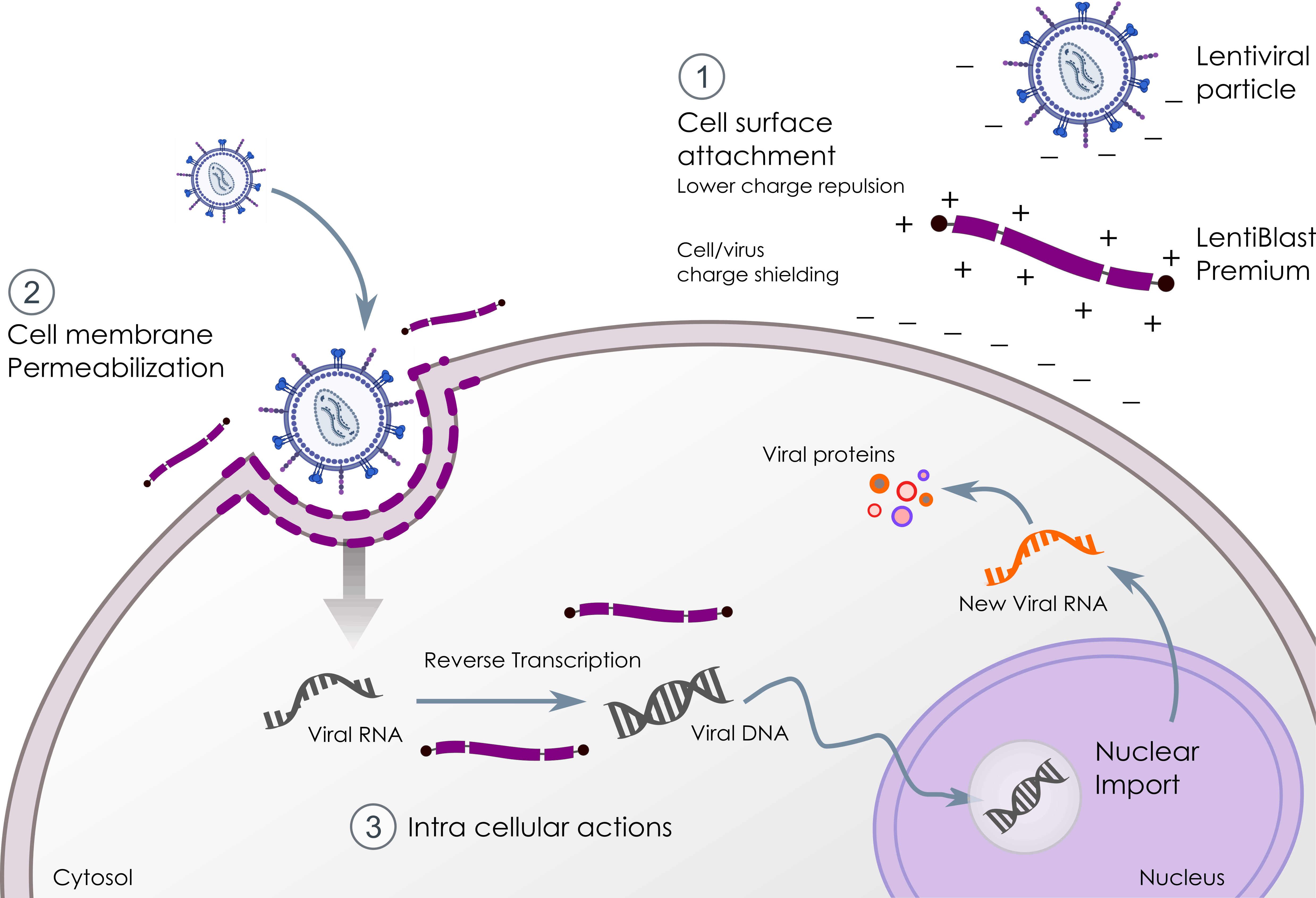
Figure 1: Schematic showing the three levels of LBP activity. (1) Cell/virus charge shielding at the surface, (2) membrane permeabilization and envelope fusion, (3) intracellular stabilization during reverse transcription and nuclear import.
Transduction of CD34+ cells with LentiBlast Premium
Dose response in the KG1a stem-cell line
Using KG1a cell line as a model of CD34+ stem cells, and a VSV-G pseudotype GFP lentivirus at a fixed MOI of 5, LBP increased transduction in a clear dose-dependent manner. Activity was measurable from as little as 0.1 µL per well and reached a plateau at 5 µL. Across the dose range, transduction rose roughly 7.5 fold, from 5.3% ± 0.5% of GFP+ cells in untreated infected controls to 40.1% ± 2.1% with LBP, confirmed independently by flow cytometry and cytofluorimetry (figure 2).
A benefit maintained across viral doses (MOI)
At a fixed 5 µL dose of LBP across MOIs from 1 to 40, the enhancer improved both the percentage of GFP+ cells and overall fluorescence intensity at every viral load. The relative benefit was greatest at low MOI, rising to a 15.1-fold increase at MOI 1 and settling to 2.8-fold at MOI 40, consistent with an apparent ceiling on the maximum transducible fraction for a given cell type.
Primary CD34+ hematopoietic stem cells
In primary CD34+ HSCs, far more sensitive than cell lines and used only at early passage, low doses of LBP (1 and 5 µL) at MOI 2 and 5 significantly increased both the fraction of GFP+ cells and fluorescence intensity. While the gain in percentage of transduced cells was more modest than in KG1a (~4-fold vs ~8-fold), the most striking effect was on fluorescence intensity, which rose from roughly 7-fold to more than 10-fold over untreated controls. Because effective cell therapy requires both a high number of transduced cells and high per-cell transgene expression, this intensity gain is clinically relevant.
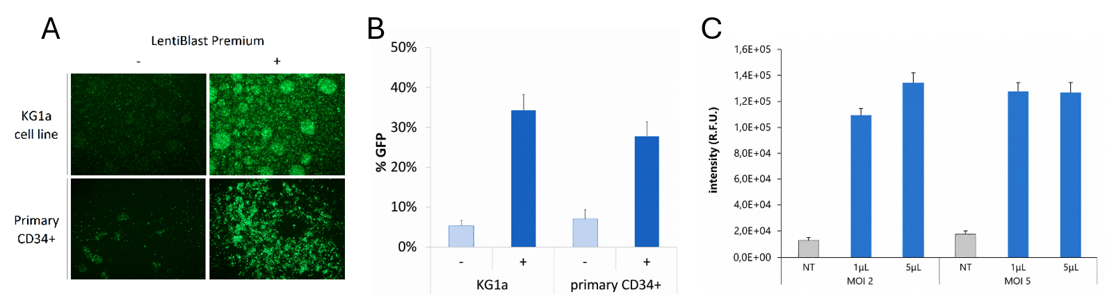
Figure 2: LBP is efficient to increase lentiviral transduction in CD34+ stem cells. GFP+ cells in CD34+ cell lines and primary cells at MOI 5 in presence or not of LentiBlast Premium monitored by fluorescence microscopy (A) or flow cytometry (B). Fluorescence intensity in primary CD34+ stem cells at MOI 2 and 5 with 1 and 5 µL LBP.
LentiBlast Premium versus Spinoculation
Spinoculation, the centrifugation of viruses onto target cells, is a widely used physical enhancement method but it is difficult to integrate into closed, bag-based manufacturing. When transducing KG1a with virus alone the overall efficiency does not exceed 5% of positive cells even at a MOI of 10. Spinoculation step only increased transduction between 2.2 and 3.9-fold depending on MOI (respectively from ~4% to 9% at MOI 10 and ~0.5% to 2% at MOI 1) and adding LBP to spinoculated cells produced no additional benefit at any MOI.
In comparison, the addition of LBP dramatically improved the lentiviral mediated transduction of KG1a at both MOI (from ~0.5% to 7.8% at MOI 1 – 15-fold, to ~4.0% to 25.8% at MOI 10 – 6.5-fold). In practical terms, KG1a transduced at a MOI of 1 in presence of 5 µL LentiBlast induced the same efficiency as spinoculated cells at 10 times more MOI without LentiBlast Premium! LBP therefore delivers spinoculation-level performance with roughly 10x less virus and no centrifugation step (figure 3).
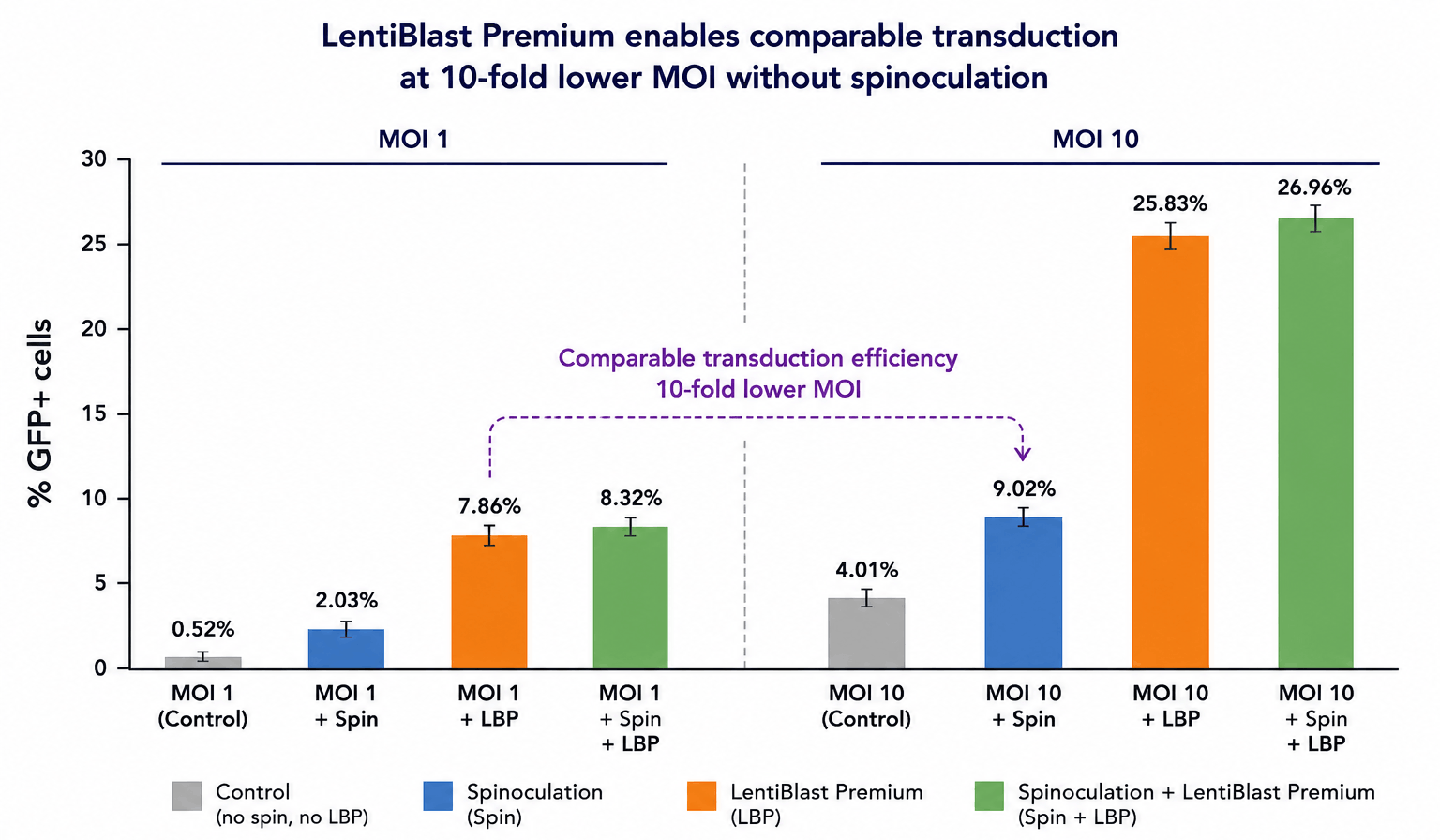
Figure 3: MOI 1 with LBP without spinoculation achieves similar transduction efficiency as MOI 10 with spinoculation KG1a were transduced with lentiviral particles at MOI 1 and 10 in the presence or absence of 5 µL LentiBlast Premium and submitted or not to spinoculation. 72H after, % of GFP+ cells were analyzed by flow cytometry.
Absence of toxicity on CD34+ cells
A transduction enhancer is only clinically useful if it preserves cell health. Across doses from 1 µL up to 20 µL per well in a 24-well plate (up to 4 times the recommended dose), LBP showed no meaningful cytotoxicity. WST-8 ranged from 100% of control at 1 µL to 84% at 20µL, and DRAQ7 staining showed no increase in late apoptosis (~10% across all conditions, including untreated). MTT analysis confirmed that any modest reduction in proliferation was attributable to the viral infection process itself rather than to LBP (table 1).
| Assay | Conditions tested | Results |
| WST-8 proliferation (72h) | Untreated, LBP 1 µL → 20 µL | 100% → 84% of control (no significant loss) |
| DRAQ7 late apoptosis | Untreated, LBP 1 µL → 20 µL | ~10% in all conditions (no increase) |
| MTT proliferation | LBP 5 µL vs virus only | No difference is attributable to LBP |
Table 1. LentiBlast Premium is non-toxic even at high doses. CD34+ cells were transduced in presence or not of ranging doses of LentiBlast Premium. Proliferation and late apoptosis were determined 72H after respectively using WST-8 assay kit, MTT assay kit or DRAQ7 staining.
Benchmark against other transduction enhancers
LentiBlast Premium was compared in KG1a against five commonly used commercial transduction enhancers (Polybrene, ViralEntry, Vectofusin-1⁴,⁵ , LentiBOOST⁶,⁷ , and Retronectin⁸) at MOI 2, 5 and 20 (figure 4). All enhancers improved transduction, but not equally and the advantage of LBP was greatest, especially at low MOI. Not only did LBP dramatically improved the transduction efficiency compared to other competitors, but also it is the most convenient to use with high viability: LBP is a ready-to-use single tube that present no toxicity even at high concentrations as opposed to Polybrene and there is no need to coat culture plate such as with Retronectin or to add cationic molecule such as with Lentiboost (table 2).
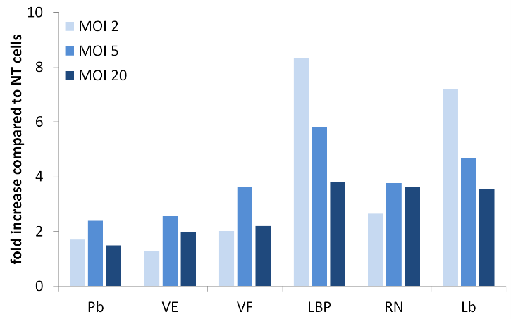
Figure 4: Fold increase in GFP+ cells over untreated controls for LBP, Polybrene (Pb), Viral entry (VE), Vectofusin-1 (VF), Retronectin (RN) and Lentiboost (Lb). CD34+ were transduced with lentiviral particles at MOI 2, 5 and 10 in the presence or absence of different viral enhancers. % of GFP+ cells were analyzed 72H after and fold increase was calculated over transduced cell without any treatment (NT).
| Attribute | LentiBlast Premium | Polybrene | Retronectin | LentiBoost | Viral Entry | Vectofusin-1 |
| Chemistry | Modified cationic poloxamer | Cationic polymer | Fibronectin fragment | Poloxamer | N/A | Amphipathic peptide |
| Fold increase @ MOI 2 | 8.3x | Modest | Modest | 7.2x | Modest | Modest |
| Efficiency at low MOI | High | Low | Low | High | Low | Low |
| Cell toxicity | None, even at high doses | High, dose dependent | Low | Possible | Moderate | Low to moderate |
| Cationic additive needed | No | N/A | No | Yes | No | No |
| Plate coating | No | No | Yes (coat + block) | No | No | No |
| Format | Single tube | Single tube | Coating reagent | Two tubes | Single tube | Single tube |
| Spinoculation required | No | Optional | Often | No | No | No |
| Magnetic-sorting compatible | Yes (ViroMICST Stem) | No | No | No | No | No |
Table 2. Head-to-head comparison of LentiBlast Premium with commonly used transduction enhancers. LentiBlast Premium combines the advantages of the other enhancers in a single reagent: a ready-to-use, single-tube formulation that needs no plate coating or spinoculation, remains non-toxic, and is compatible with magnetic cell sorting.
ViroMICST Stem: adding magnetic guidance
Classical poloxamers are non-ionic and destabilize when combined with magnetic nanoparticles, causing precipitation, loss of efficiency and toxicity. Because LBP carries cationic groups, it can be formulated with a dedicated magnetic-beads formulation, ViroMICST Stem (VMStem), making the chemistry amenable to Magnetofection and compatible with closed immuno-magnetic cell sorting systems (figure 5).
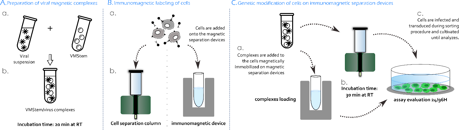
Figure 5: Protocol for ViroMICST Stem genetic modification during immunomagnetic cell sorting. (A) Complexes are prepared by mixing viral suspension with ViroMICST Stem. (B) After immunomagnetic staining, the cells are loaded onto the magnetic separation device. Magnetic complexes are then added to the cells retained and incubated 30 min at room temperature (C) before being cultivated until evaluation of the experiment.
Genetic modification during immunomagnetic cell sorting
Building on the earlier Integrated Magnetic Immuno-Cell Sorting and Transfection/Transduction (iMICST) technology concept⁹, VMStem enables an original protocol in which target cells are transduced while immobilized on an immunomagnetic separation device (figure 6). Magnetically labelled KG1a cells are loaded onto an MS column or an EasySep magnet; magnetized viral complexes were then added and transduced the retained cells during a 30-minute incubation. On the MS column, adding VMStem to immobilized cells raised transduction from 3.3% ± 1.0% to 37.5% ± 2.3% (an 11.5-fold increase), and comparable gains were obtained on the EasySep magnet (~30% GFP+ cells, 9.6-fold) (table 3).
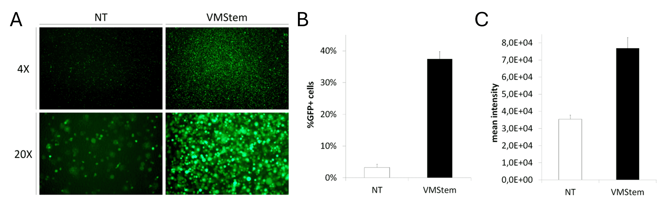
Figure 6: ViroMICST Stem (VMStem) improves viral transduction on immunomagnetic separation devices. (A) representative GFP fluorescence images following viral transduction on immunomagnetic separation device in the absence (NT) or presence of VMStem (4X and 20x). (B) Percentage of GFP+ cells and (C) mean GFP fluorescence intensity were measured by flow cytometry 72H after transduction.
| Condition (KG1a, MOI 5, 72H incubation) | GFP+ cells | Fold vs untreated |
| MS Column, virus only | 3.3% ± 1.0% | N/A |
| MS Column + ViroMICST Stem | 37.5% ± 2.3% | 11.5x |
| EasySep magnet + ViroMICST Stem | ~30% | 9.6x |
Table 3. Transduction of CD34+ cells directly on immunomagnetic sorting devices using ViroMICST Stem. Genetic modification occurs during the purification step itself. Immunomagnetically labelled cells come into contact with magnetized viral particles and get transduced while immobilized into the column by the separation magnet.
Practical and economic benefits
A single-tube protocol, no coating or spinoculation needed
LentiBlast Premiu is added directly to the cells together with the viral vector: no plate coating, no blocking, no spinoculation and no medium change required before or after transduction. It is supplied as a single tube, in contrast to two-component enhancers that must be optimized against each other. This simplicity reduces handling, shortens the workflow and lowers the risk of process variability.
Cell viability
This new class of patented cationic poloxamers showed no detectable cytotoxicity under tested conditions and is compatible with cell life and it allows to perform transduction under mild conditions as it does not require prolonged incubation or additional manipulation steps. Cells maintain high viability and functional properties following the procedure. This makes LentiBlast Premium and the magnetic formulations that derive (ViroMICST Stem) particularly suitable for sensitive primary hematopoietic stem cells and progenitor cells intended for downstream culture of therapeutic applications.
Less virus, lower cost per dose
Because viral production is the dominant cost driver in cell therapy manufacturing, achieving equivalent transduction at low MOI translates directly into reduced vector consumption per dose, with downstream benefits for cost of goods and, ultimately, patient access. The spinoculation comparison above illustrates the magnitude: LBP delivered comparable transduction with roughly 10x less virus.
Market context. Viral vector supply is the single largest controllable cost in ex vivo gene-modified cell therapy manufacturing. Industry estimates place GMP lentiviral vector production at up to 40% of the total cost of goods, within a total manufacturing cost of goods commonly estimated at $0.5 to 1 million per patient. Expressed per batch, a single patient-scale viral batch can cost in excess of $16,000¹⁰. The same economics are reflected in the price of approved autologous lentiviral HSC therapies, from $2.8 million for Zynteglo and $3.0 million for Skysona to $3.1 million for Lyfgenia and $4.25 million for Lenmeldy (Libmeldy in Europe), currently the most expensive medicine worldwide. Any reagent that achieves equivalent transduction at a lower MOI therefore acts directly on the main cost lever of the process, and on the affordability that conditions patient access.
Compatible with automated, closed systems
LBP and VMStem are designed with clinical manufacturing in mind. VMStem is compatible with immunomagnetic separation columns and magnets, and the on-device transduction protocol is one step away from fully automated instruments such as the CliniMACS Prodigy®, which is approved for clinical use and enables reproducible, GMP-grade CD34+ processing. A GMP grade of LentiBlast Premium is in development.
Where LentiBlast Premium fits
LentiBlast Premium was developed for lentiviral and retroviral gene transfer into hard-to-transduce primary cells, with CD34⁺ hematopoietic stem cells as the primary target. Since its launch it has been cited in around one hundred peer-reviewed publications across a wide range of cell types and experimental models.
HSC gene therapy. The core application is ex vivo gene transfer into CD34⁺ cells for autologous transplantation, the manufacturing route behind approved and investigational therapies for β-thalassemia, sickle cell disease, metachromatic leukodystrophy and cerebral adrenoleukodystrophy. LBP delivers higher transduction at lower MOI while preserving viability and phenotype, in a workflow compatible with closed systems. Its value has been demonstrated in vivo: mouse hematopoietic stem cells transduced with a SUMF1 lentiviral vector in the presence of LBP and transplanted into recipient mice corrected the underlying biochemical deficit and improved neurocognitive function¹¹.
CAR-T and immune cell engineering. Primary human T cells transduced with a CAR-encoding lentiviral vector in the presence of LentiBlast Premium reached more than 96% CAR-positive cells at day 7¹, a level directly relevant to CAR-T manufacturing. Because LBP requires no plate coating and works in any culture vessel, from multiwell plates to culture bags, it inserts into existing T cell workflows without process redesign.
Genetic modification during immunomagnetic sorting. Through ViroMICST Stem, purification and genetic modification are collapsed into a single step performed on the separation column or magnet, saving time, reducing handling and cutting viral vector consumption. This is the step that brings the process closest to fully automated, closed platforms.
Preclinical research and disease modelling. LBP has enabled experiments that are difficult to perform with conventional enhancers, including the transduction of primary acute myeloid leukemia blasts from patients, where 30 to 60% efficiency allowed the functional consequences of RNA splicing factor mutations to be dissected, and the generation of retrogenic TCR mouse models from retrovirally transduced hematopoietic stem cells¹². Reported applications also include primary myeloid precursors, cardiomyocytes and trabecular meshwork cells.
Beyond lentiviral vectors. The same modified poloxamer chemistry has been extended to AAV with AAVBlast, a cationic poloxamer formulation characterised in a recent peer-reviewed study co-authored by OZ Biosciences¹³. Screened against a panel of poloxamers, AAVBlast was identified as the most effective enhancer, producing a 6.4-fold increase in transgene expression in primary ovine mesenchymal stromal cells, with comparable gains in HeLa, HEK293T and HUVEC cells and across the AAV2, AAV6 and AAV8 serotypes. Mechanistically, it stabilizes viral particles and increases their bioavailability: detectable capsid levels rose 6.2-fold after incubation at 37 °C, and treatment significantly increased intracellular AAV DNA, transgene mRNA and protein output in every cell type tested. Notably, AAVBlast did not enhance transduction of primary monocyte-derived macrophages, indicating that its activity is confined to cells naturally permissive to AAV and that the favourable immunological profile of AAV vectors is preserved. Applied to a chitosan/β-tricalcium phosphate gene-activated matrix, AAVBlast increased the recovery of functional vector 4.2-fold and raised BMP-2 and VEGF secretion 2.29- and 1.76-fold respectively, extending the platform from ex vivo cell manufacturing to localised gene delivery for tissue regeneration.
Product grades. LentiBlast Premium is available in a research grade and in a Superior Grade, identical in synthesis and formulation but with additional characterisation and purity testing, meeting USP ⟨63⟩ for mycoplasma and USP ⟨85⟩ for bacterial endotoxins. The Superior Grade is intended for preclinical and early phase clinical use as an ancillary material for cell and gene therapy. A GMP grade is in development.
Summary
Transduction efficiency is the rate-limiting step that determines the feasibility, cost and clinical scalability of HSC gene therapy. The difficulty of transducing CD34+ stem cells at high efficiency and low MOI has imposed constraints on manufacturing economics and safety that conventional enhancers cannot fully overcome.
LentiBlast Premium addresses these limitations. As a new class of cationic poloxamer, it delivers strong, dose-dependent transduction enhancement in both CD34+ cell line and primary HSCs while remaining non-toxic even at twice the recommended dose, requiring no cationic additive, and eliminating spinoculation and plate coating. It matches spinoculation performance with roughly 10x less virus and outperforms leading enhancers at low MOI, where transduction is hardest to achieve. Through ViroMICST Stem, it uniquely enables genetic modification of cells directly on immunomagnetic sorting devices, and supports closed-system, automated HSC manufacturing.
The benefits apply broadly across cell and gene therapy: reduced viral vector consumption, lower cost of goods, a simpler and safer workflow, and improved access to affordable curative therapies. With GMP grades in development, LentiBlast Premium and ViroMICST Stem help lower the barriers to efficient and affordable gene therapy.
Author: Cédric Sapet, PhD.
References
¹ Sta Maria NS, et al. Sci Rep. 2021 Jul 23;11(1):15077.
² Finkelshtein D, et al. LDL receptor and its family members serve as the cellular receptors for vesicular stomatitis virus. Proc Natl Acad Sci USA. 2013;110(18):7306-7311.
³ Ginn SL, et al. Gene therapy clinical trials worldwide to 2023: an update. J Gene Med. 2024;26:e3721.
⁴ Fenard D, et al. Vectofusin-1, a new viral entry enhancer, strongly promotes lentiviral transduction of human hematopoietic stem cells. Mol Ther Nucleic Acids. 2013;2:e90.
⁵ Majdoul S, et al. Molecular determinants of Vectofusin-1 and its derivatives for the enhancement of lentivirally mediated gene transfer into hematopoietic stem/progenitor cells. J Biol Chem. 2016;291(5):2161-2169.
⁶ Hauber I, et al. Improving lentiviral transduction of CD34+ hematopoietic stem and progenitor cells. Hum Gene Ther Methods. 2018;29(2):104-113.
⁷ Uchida N, et al. High-efficiency lentiviral transduction of human CD34+ cells in high-density culture with poloxamer and prostaglandin E2. Mol Ther Methods Clin Dev. 2019;13:187-196.
⁸ Hanenberg H, et al. Colocalization of retrovirus and target cells on specific fibronectin fragments increases genetic transduction of mammalian cells. Nat Med. 1996;2(8):876-882.
⁹ Sanchez-Antequera Y, et al. Magselectofection: an integrated method of nanomagnetic separation and genetic modification of target cells. Blood. 2011;117(16):e171-e181.
¹⁰ Abdo L, et al. Cost-effective strategies for CAR-T cell therapy manufacturing. Mol Ther Oncol. 2025; 33(2):200980.
¹¹ Pham V, et al. Hematopoietic stem cell gene therapy improves outcomes in a clinically relevant mouse model of multiple sulfatase deficiency. Mol Ther. 2024; 32(11):3829-3846.
¹² Burton OT, et al. The tissue-resident regulatory T cell pool is shaped by transient multi-tissue migration and a conserved residency program. Immunity. 2024;57(7):1586-1602.
¹³ Musoki A, et al. Development of a gene-activated matrix for enhanced AAV gene delivery in vitro. Front Bioeng Biotechnol. 2026;10:14:1832901.
Related Articles

Cytokine-fusion scaffold generates CAR T cells enriched in long-lived TSCM

Enhancing transduction efficiency of lentiviral vectors in CAR-T cell therapy