





BLOG > Publications & Citations > NGF cmRNA-engineered stem cells induce functional neurite outgrowth
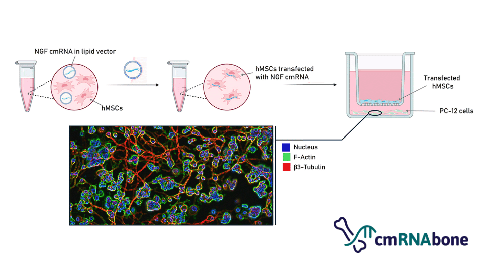
Paper from Polain C et al., mRNA-Engineered Stem Cells Produce Nerve Growth Factor and Induce Neurite Outgrowth in Recipient Cells. Exp Cell Res. 2026 Sep 28;463(1):115188.
This study was carried out within the framework of the cmRNAbone project (EU Horizon 2020, grant No. 874790), which aims to develop chemically modified mRNA as a next-generation platform for bone repair.
Introduction
Bone is not just mineral and collagen, it is a highly innervated tissue, and innervation plays a critical role in bone healing. Yet this dimension has been largely overlooked in regenerative medicine. Polain et al. set out to change that by engineering human bone marrow-derived mesenchymal stem cells (hMSCs) with chemically modified mRNA (cmRNA) encoding nerve growth factor (NGF) and showing for the first time that this transcript-based approach is sufficient to induce functional neurite outgrowth in recipient neural cells in vitro. Central to this work: all lipid vectors used were developed and provided by OZ Biosciences.
Key results
This study produced four interconnected findings that establish NGF cmRNA therapy as a credible neurotrophic strategy for bone regeneration:
- A transcript-based approach matches recombinant protein, at 4.5x lower concentrations. hMSCs transfected with NGF cmRNA produced peak NGF levels of ~8 ng/ml at day 1 post-transfection, driving neurite outgrowth in PC-12 recipient cells comparable to that of direct stimulation with recombinant human NGF (rhNGF) at 50 ng/ml. This means that endogenously produced NGF was therapeutically effective at concentrations 4.5 times lower than the recombinant protein demonstrating a key advantage for clinical translation.
- NL37 outperforms all tested formulations in hMSCs, including Lipofectamine™ MessengerMax™. Among five lipid vectors evaluated, the novel OZ Biosciences formulation NL37 (used at a 1:4 cmRNA-to-lipid ratio) delivered the highest and most sustained MetLuc and NGF cmRNA expression in primary hMSCs, while maintaining cell viability above 75%. Other lipid such as NL51 failed to match these results due to significant toxicity (values <66%, p <0.0001).
- NGF cmRNA rewires the stem cell proteome toward neurogenesis. Proteomic analysis of NGF cmRNA-transfected hMSCs revealed 115 significantly upregulated proteins, including neurogenesis-associated factors such as β3-Tubulin, vitronectin (VTN, 5.54-fold), and apolipoproteins APOB-100 and APOE (>3.8-fold). These data suggest that beyond serving as protein factories, hMSCs transfected with NGF cmRNA begin to acquire a neurogenic-like molecular identity.
- PC-12 recipient cells show robust, dose-dependent neurite outgrowth. In the co-culture system, PC-12 cells co-cultured with NGF cmRNA-transfected hMSCs showed a steady, significant increase in total neurite length, neurite area, and branch points from day 2 onwards (p <0.0001). NL37 formulations at 31.2 pg cmRNA/cell achieved the highest total neurite length (3.80 ± 0.88 cm, p <0.05) at day 5, significantly outperforming Lipofectamine MessengerMax at the same dose.
Methodology: a staged, multi-platform screening strategy
The study followed a structured, multi-step approach designed to progressively identify optimal conditions before advancing to the biological readout:
Stage 1 - High-throughput screening in A549 cells.
Five lipid vectors all developed by OZ Biosciences (RmesFect™, 3D-FectIN™, 3D-Fect™, NL51, and NL37) were evaluated across 12 cmRNA-to-lipid ratios (1:0.5 to 1:11) and 7 cmRNA concentrations (0.78 to 50 pg/cell) using MetLuc cmRNA as a reporter. Protein expression and cell viability over 3 days provided a preliminary ranking of formulations.
Key finding: cmRNA concentrations >25 pg/cell were cytotoxic regardless of lipid, while NL37 and NL51 at 1:4 consistently delivered the highest expression with acceptable viability (Fig. 1).
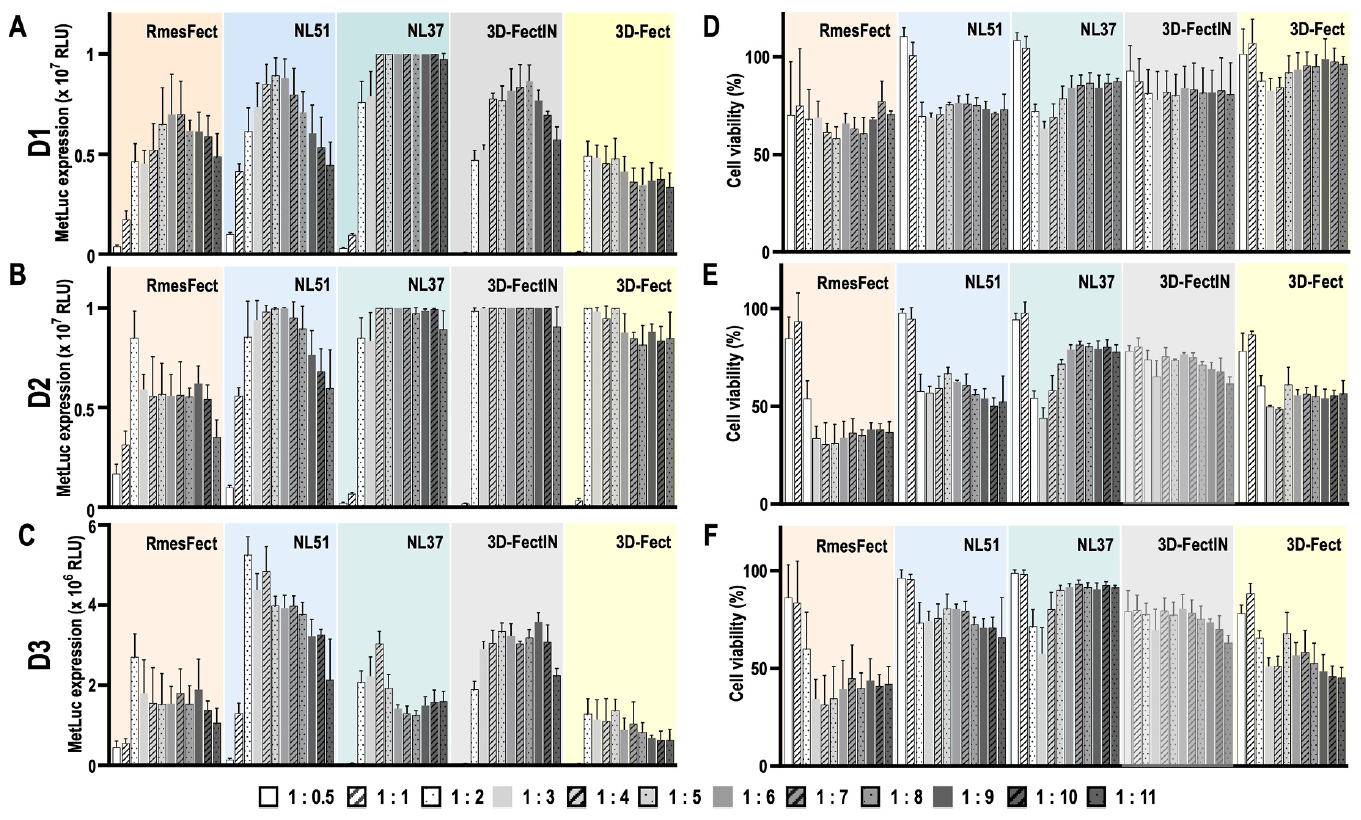
Figure 1: Protein expression and biocompatibility in A549 cells are dependent on the cmRNA-to-lipid ratio. (A-C) MetLuc expression at days 1, 2, and 3 post-transfection across five OZ Biosciences lipid vectors and 11 cmRNA-to-lipid ratios. (D-F) Corresponding cell viability values. NL37 at 1:4 emerged as the best formulation, combining high protein expression with maintained cell viability. Adapted from Polain et al., Exp. Cell Res. 2026, 115188.
Among all the conditions tested, NL37 and NL51 showed the highest protein expression over 3 days at a 1:4 cmRNA-to-lipid ratio, and were both selected alongside RmesFect™ for further evaluation in primary hMSCs.
Stage 2 - Direct evaluation in primary hMSCs.
Selected conditions were translated to primary bone marrow-derived hMSCs using MetLuc, eGFP, and NGF cmRNA. RmesFect™, NL51, and NL37 were tested at the ratios identified in Stage 1. NL37 at 1:4 delivered superior MetLuc expression over 5 days (p <0.0001 vs. NL51 and RmesFect™) while maintaining cell viability ≥75%, confirming it as the formulation of choice for subsequent NGF experiments (Fig. 2).
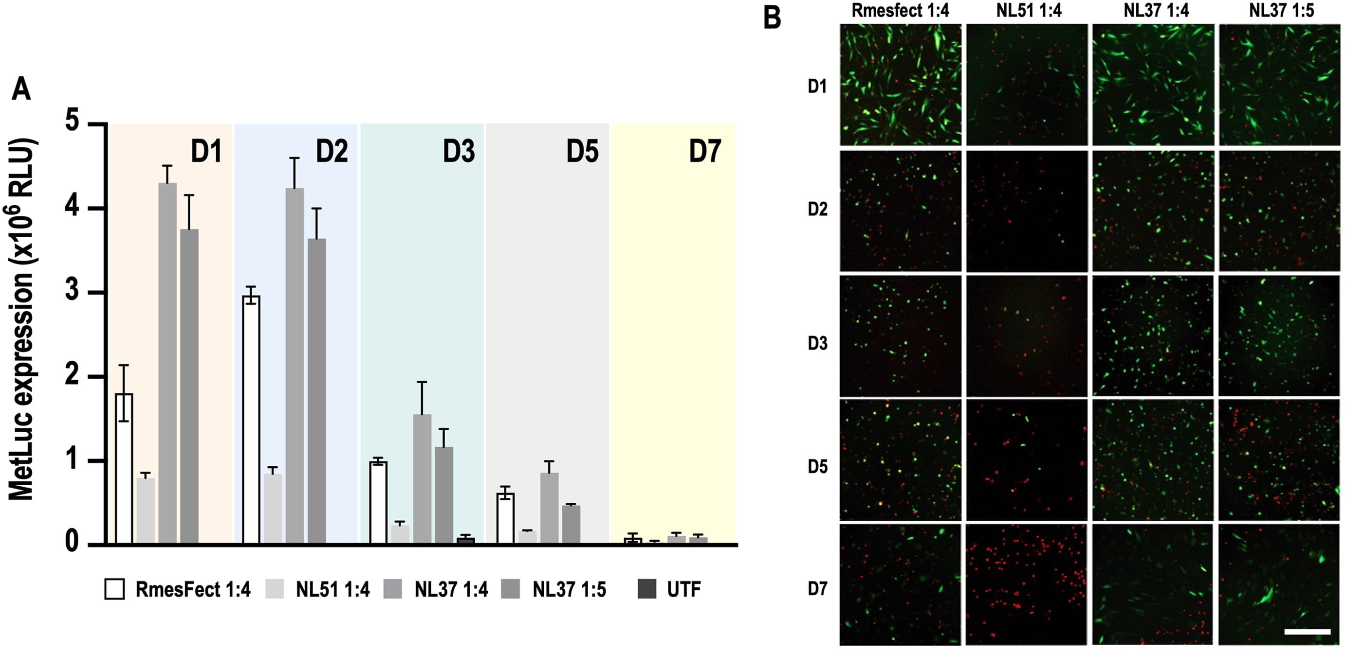
Figure 2: NL37 stands up as the most suitable lipid vector for cmRNA transfer to hMSCs. (A) MetLuc expression in hMSCs transfected with RmesFect™, NL51, NL37 1:4, and NL37 1:5 over 7 days. NL37 1:4 showed the highest and most sustained expression. (B) eGFP fluorescence imaging confirmed transfection efficiency across conditions, with NL51 showing clear cytotoxicity (red dead cells). Adapted from Polain et al., Exp. Cell Res. 2026, 115188.
Stage 3 - NGF cmRNA transfection in suspension for co-culture.
hMSCs were transfected in suspension using NL37 (1:4) and Lipofectamine MessengerMax across four NGF cmRNA concentrations (15.6, 31.2, 62.5, and 100 pg/cell). Transfection in suspension significantly increased protein expression vs. adherent monolayers (8.4 × 10⁶ RLU at day 1, p <0.0001). NGF concentrations were measured by ELISA daily for 7 days, and transfected hMSCs were immediately placed in co-culture with PC-12 cells on laminin/poly-D-lysine coated coverslips.
Stage 4 - Co-culture and neurite quantification.
A semipermeable insert system (1 µm TRAKETCH® membrane) separated hMSCs from PC-12 cells, allowing paracrine NGF signaling without direct contact. Neurite length, area, and branch points were quantified by automated image analysis (NIS-Elements GA3) over 7 days. NGF secretion by hMSCs in co-culture was measured by ELISA (Fig. 3), while neurite outgrowth in PC-12 was assessed by fluorescence microscopy (Fig. 4).
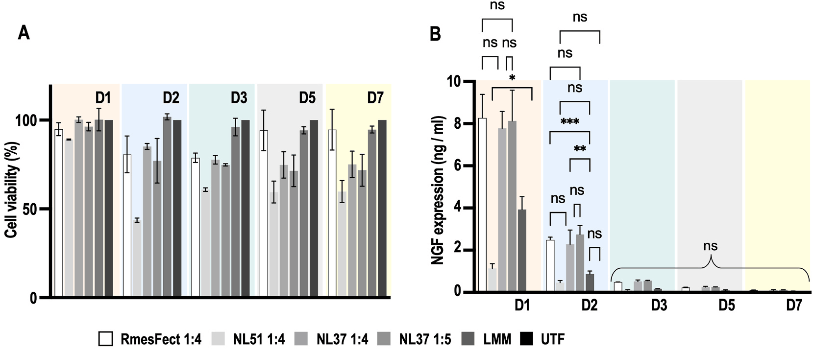
Figure 3: NGF cmRNA-transfected hMSCs secrete NGF in vitro and activate neurogenesis-related proteomic pathways. (A) Cell viability across lipid formulations over 7 days - NL51 showed significant toxicity (values <66%, p <0.0001); RmesFect™ and NL37 maintained viability ≥75%. (B) NGF secretion kinetics measured by ELISA: RmesFect™ and NL37 reached ~8 ng/ml at day 1, significantly higher than Lipofectamine MessengerMax (3.9 ng/ml, p <0.0001). Adapted from Polain et al., Exp. Cell Res. 2026, 115188.
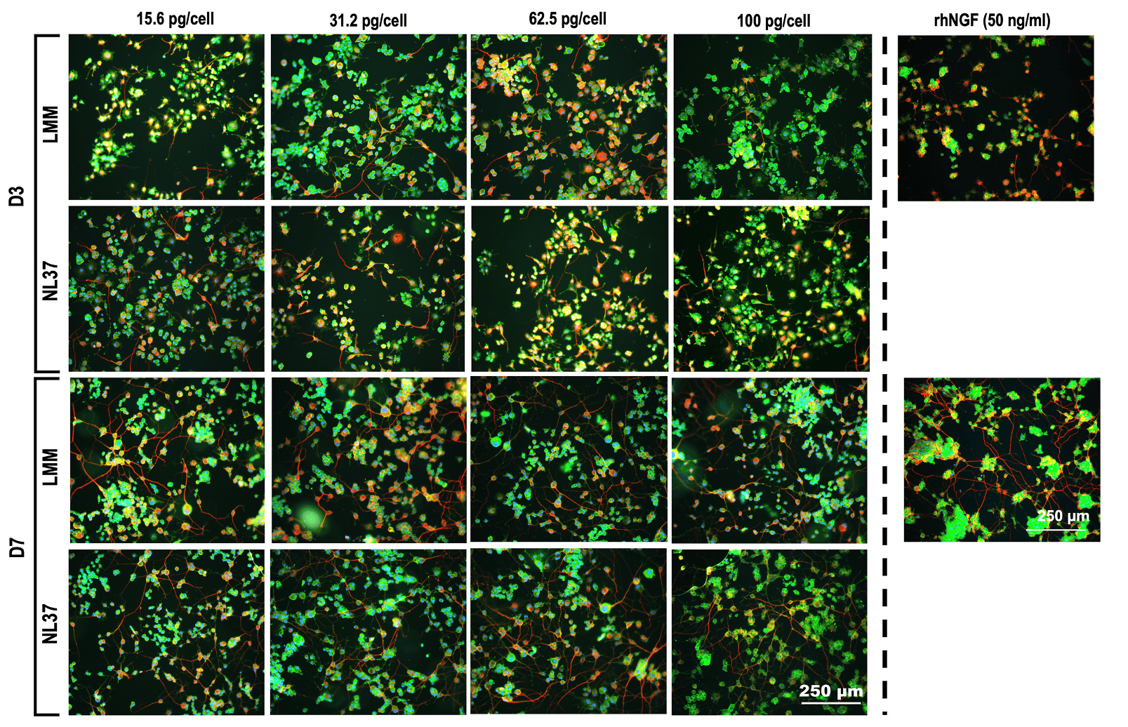
Figure 4: Fluorescence micrographs of PC-12 cells after co-culture with NGF cmRNA-transfected hMSCs (day 3 and day 7). Blue: nuclei, green: F-Actin, red: β3-Tubulin. Four cmRNA concentrations (15.6, 31.2, 62.5, and 100 pg/cell) with NL37 or Lipofectamine MessengerMax (LMM) are shown alongside recombinant human NGF (50 ng/ml) as positive control. Dense neurite networks are clearly visible by day 7 with NL37. Scale bar: 250 µm. Adapted from Polain et al., Exp. Cell Res. 2026, 115188.
These results confirmed that NGF produced by cmRNA-transfected hMSCs is biologically active and sufficient to drive neurite outgrowth in recipient PC-12 cells, with NL37 consistently outperforming Lipofectamine MessengerMax across all conditions tested.
OZ Biosciences Lipid Vectors in This Study
Five lipid vectors developed and provided by OZ Biosciences (Marseille, France) were central to this study: RmesFect™, 3D-FectIN™, 3D-Fect™, NL51, and the novel experimental formulation NL37. All are patented molecules (US9107931B2) sharing a common structural backbone: a hydrophobic unsaturated tail, a biocompatible linker, and a polar head based on basic natural amino acid residues, but differing in polar head charge and lipid-to-co-lipid ratio, properties that critically determine their transfection efficiency and biocompatibility in a cell-type-specific manner.
In the initial A549 screen, NL37, NL51, and 3D-FectIN™ reached the highest MetLuc expression values (1.64-1.65 × 10⁷), significantly outperforming RmesFect™ and 3D-Fect™ (p <0.0001). However, cell-type specificity proved decisive: NL51, which performed comparably to NL37 in A549, showed markedly reduced efficiency and significant toxicity in primary hMSCs: an important reminder that lipid screening in cell lines cannot substitute for validation in primary target cells.
NL37 distinguished itself in hMSCs through a unique structural feature: an excess of cationic lipid relative to co-lipid, and the formation of distinctive "bleb"-structured lipoplex particles, a morphology recently associated with enhanced mRNA integrity and transfection potency. Used at a 1:4 cmRNA-to-lipid ratio and 15.6-62.5 pg cmRNA/cell, NL37 delivered the highest sustained NGF expression and neurite outgrowth parameters across all conditions tested, making it the lipid of choice for this NGF cmRNA co-culture platform. Its performance in suspension transfection, a more demanding format, further confirmed its versatility and robustness (Fig. 5).

Figure 5: Transmission electron microscopy (TEM) micrographs of cmRNA lipoplexes formed with each OZ Biosciences lipid vector and Lipofectamine MessengerMax (LMM), using NGF cmRNA at a 1:4 RNA-to-lipid ratio. The distinctive "bleb" structure of NL37 lipoplexes is indicated by red arrows - a morphology recently associated with enhanced mRNA integrity and transfection potency. Scale bar: 50 nm. Adapted from Polain et al., Exp. Cell Res. 2026, 115188.
NL37 vs Lipofectamine MessengerMax: a head-to-head comparison
Lipofectamine MessengerMax was included in this study as a reference standard, representing the current gold-standard for mRNA transfection in the field. The data consistently favored NL37 across every performance dimension evaluated.
In terms of NGF secretion, RmesFect™ and NL37 achieved peak levels of approximately 8 ng/ml at day 1 in hMSC monoculture, significantly higher than the 3.9 ng/ml produced by Lipofectamine MessengerMax (p <0.0001). In the co-culture setting, NL37 at 62.5 pg cmRNA/cell produced 11.2 ng/ml of secreted NGF at day 2, outperforming Lipofectamine MessengerMax at 100 pg cmRNA/cell (9.4 ng/ml), meaning NL37 required 1.6× less RNA to achieve a superior biological output (Fig 6). In terms of neurite outgrowth, NL37 complexes at 31.2 pg cmRNA/cell were significantly better than Lipofectamine MessengerMax at the same dose from day 3 onwards (p <0.04), with total neurite length reaching 3.80 ± 0.88 cm at day 5 vs. lower values for LMM. Finally, at the physicochemical level, NL37 complexed 94.63% of the available cmRNA vs. 74.97% for Lipofectamine MessengerMax - a 27% gain in complexation efficiency that directly translates into more biologically active material delivered per dose.
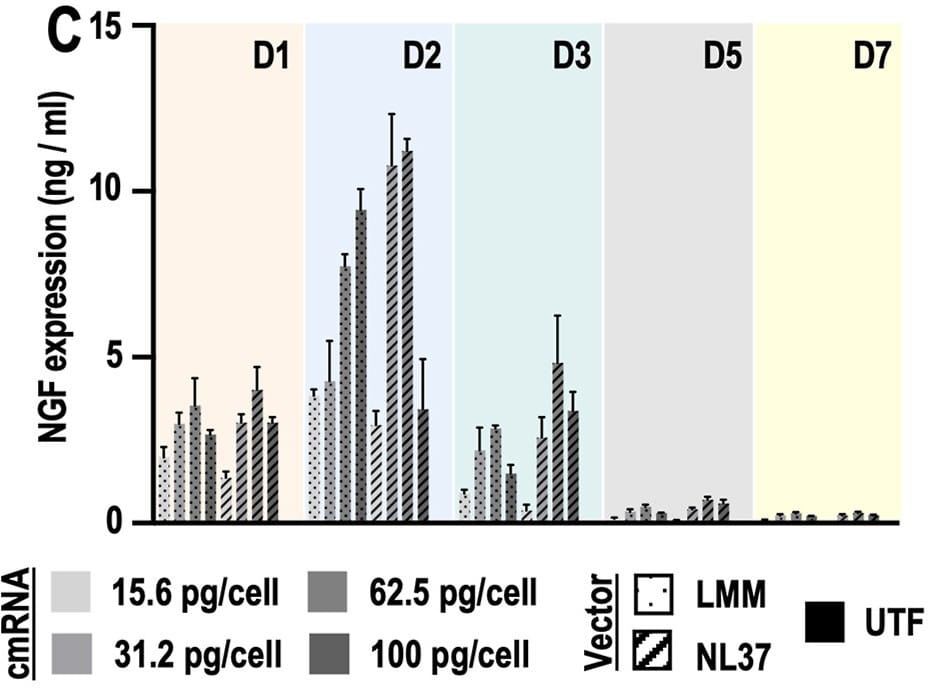
Figure 6: NGF secretion kinetics in the co-culture setting over 7 days. NL37 at 62.5 pg/cell achieved 11.2 ng/ml at day 2, outperforming Lipofectamine MessengerMax at 100 pg/cell (9.4 ng/ml). Adapted from Polain et al., Exp. Cell Res. 2026, 115188.
Link to the cmRNAbone Grant Project - Horizon 2020
This work was conducted within the framework of the cmRNAbone project, funded by the European Union's Horizon 2020 research and innovation programme under grant agreement No. 874790. cmRNAbone is dedicated to harnessing chemically modified mRNA as a next-generation platform for bone repair, exploring the combination of osteogenic, angiogenic, and now neurotrophic cmRNA signals to recreate the full regenerative microenvironment of healing bone tissue. This publication adds an important neurotrophic dimension to the project's therapeutic arsenal, laying the groundwork for future multi-signal cmRNA strategies targeting critical-sized bone defects.
Reference
Polain C, Del Toro Runzer C, Bouwman FG, Rademakers T, Haartmans M, Peniche Silva CJ, Bonvin E, Poulhes F, Plank C, Cillero-Pastor B, van Griensven M and Balmayor ER. mRNA-Engineered Stem Cells Produce Nerve Growth Factor and Induce Neurite Outgrowth in Recipient Cells. Experimental Cell Research, Volume 463, Issue 1, 2026, 115188, ISSN 0014-4827. doi: 10.1016/j.yexcr.2026.115188
Related Product
- RmesFect™: Lipid vector specifically designed for mRNA delivery, suitable for adherent cell monolayers including primary MSCs.
- 3D-FectIN™: Versatile lipid vector for mRNA transfection in 3D culture conditions and suspension formats.
- 3D-Fect™: Optimized for mRNA delivery in 3D systems with a higher positive charge density than RmesFect™.
- NL51 / NL37: Novel experimental cationic lipid formulations from OZ Biosciences, part of a next-generation series with unique structural features (bleb-forming NL37) that can deliver superior transfection in specific cell types, including primary hMSCs. Contact OZ Biosciences for availability.
All lipid vectors are part of OZ Biosciences' patented mRNA delivery platform (US9107931B2).
Related publications

Development of a gene-activated matrix for enhanced AAV gene delivery in vitro
Circulating miRNAs are associated with successful bone regeneration
Geneformer-guided multiomics identifies Pbx1 as a network hub of HSC aging